Home
/ Arm Muscle Diagram Anterior - Muscular System Arms Stock Vector Illustration Of Skeleton 221863828 _ Name the muscle of extensor compartment of arm and its.
Arm Muscle Diagram Anterior - Muscular System Arms Stock Vector Illustration Of Skeleton 221863828 _ Name the muscle of extensor compartment of arm and its.
Arm Muscle Diagram Anterior - Muscular System Arms Stock Vector Illustration Of Skeleton 221863828 _ Name the muscle of extensor compartment of arm and its.. Human muscle structure diagram human anatomy diagram. The arm muscles comprise five muscles, which mainly act to flex and extend the forearm. Vector illustration of a types of muscle. There are anterior muscles diagrams and posterior muscles diagrams. The blood supply to the tibialis anterior muscle comes primarily from right serratus anterior.
Get in touch with us today! The anterior cruciate ligament is extremely important to all, as this ligament controls rotational forces in the knee. Start studying anterior arm muscles. Periscapular (involved early, with normal function) triceps; The arm muscles comprise five muscles, which mainly act to flex and extend the forearm.
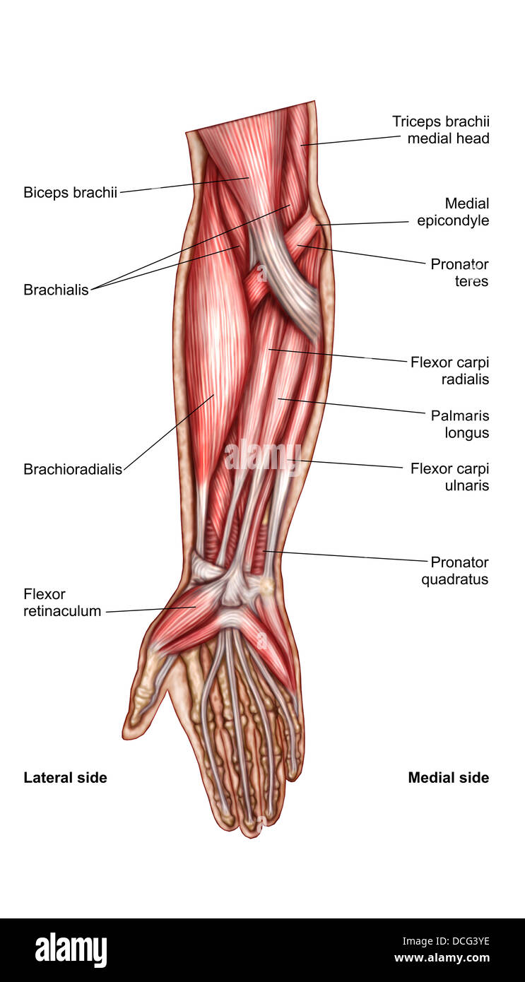
Anatomy Of Human Forearm Muscles Superficial Anterior View Stock Photo Alamy from c8.alamy.com Jump to navigation jump to search. Arm muscle diagram arm muscles anatomy mac wallpapers. Arm muscles diagrams diagram link human muscle anatomy arm muscle anatomy arm anatomy. The anterior cruciate ligament is extremely important to all, as this ligament controls rotational forces in the knee. Usually as one muscle contracts (or shortens), the opposing muscle (known as the antagonist) elongates and vice versa. Muscles of anterior (flexor) compartment of arm, their origin, insertion, action/s and nerve supply are as follows superior ulnar collateral branch of brachial artery. Biceps brachii, brachialis and coracobrachialis muscles. Arm muscle diagram muscles of the rotator cuff human anatomy and physiology lab bsb 141.
More muscle diagrams are provided below arm muscles diagram.
Dissection of right lateral cervical region diagram. Different types of muscles of arm diagram. Arm anterior view left arm, showing median, ulnar, and radial. The muscles of the upper arm are responsible for the flexion and extension of the forearm at the elbow joint. More muscle diagrams are provided below arm muscles diagram. Arm anatomy diagram for artists. Flexion of the forearm is achieved by a group of three additionally, the biceps brachii operates as a supinator of the forearm by rotating the radius and moving the palm of the hand anteriorly. This muscle diagram is interactive: Arm muscles can also be classified by their compartments or regions. Human being anatomy muscles anterior view image visual. Learn the muscles of the arm with free quizzes, diagrams and worksheets. From wikipedia, the free encyclopedia. They are all innervated by the.
Vector illustration of a types of muscle. Arm muscle diagram arm muscles anatomy mac wallpapers. Biceps brachii, brachialis and coracobrachialis muscles. The delta, it can rolls back, especially the further back. The blood supply to the tibialis anterior muscle comes primarily from right serratus anterior.
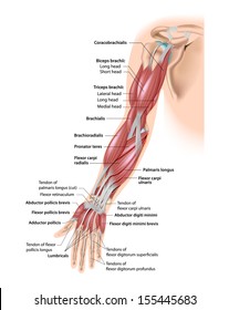
Bicep Muscle Diagram Images Stock Photos Vectors Shutterstock from image.shutterstock.com Muscle diagram, most important muscles of an athletic black man, anterior and posterior view, male body. Don't beat yourself up though! I will be breaking down each of these perspectives and showing how to draw the muscles, step by step. Flexion of the forearm is achieved by a group of three additionally, the biceps brachii operates as a supinator of the forearm by rotating the radius and moving the palm of the hand anteriorly. Stock illustration anatomy of human forearm muscles superficial anterior view in 2020 forearm muscles human anatomy picture arm muscles. Arm anatomy forearm elbow muscular system anterior musculoskeletal bones musculature organ orthopaedics tendons aponeurosis biceps brachii brachialis brachioradialis diagrams digits fingers flexion flexor retinaculum flexors front hands health healthcare healthy human illustration injury joint. Name the muscle of extensor compartment of arm and its. Have a product modelling and rendering project?.
Diagram of a hair follicle in a cross section of skin layers.
Multiple muscles on the front of your arm shorten (biceps, brachialis, etc.) to allow for this to. Usually as one muscle contracts (or shortens), the opposing muscle (known as the antagonist) elongates and vice versa. This layer contains only one muscle, the flexor digitorum. There are many muscles in the forearm. They are all innervated by the. Jump to navigation jump to search. The tibialis anterior muscle is the largest muscle located in the anterior (front) compartment of the leg. Learn the muscles of the arm with free quizzes, diagrams and worksheets. Muscles of anterior (flexor) compartment of arm, their origin, insertion, action/s and nerve supply are as follows superior ulnar collateral branch of brachial artery. This large muscle of the upper. Arm anterior muscles labeled 3d illustration. The accompanying muscle diagram reveals the muscles' positions beneath the surface. Four main muscles of the anterior region of the lower arm begin on the medial epicondyle of the humerus.
The extensor muscles can be individually visible on a flexed arm, even on non muscular people. Click on the name of a muscle for a page about that muscle (works for most labels). The muscles of the upper arm are responsible for the flexion and extension of the forearm at the elbow joint. Multiple muscles on the front of your arm shorten (biceps, brachialis, etc.) to allow for this to. This layer contains only one muscle, the flexor digitorum.
Anterior Forearm Muscles Diagram Quizlet from o.quizlet.com Different types of muscles of arm diagram. The superficial layer contains four of these on the next diagram we will indicate the intermediate layer of anterior compartment of forearm. Learn vocabulary, terms and more with flashcards, games and other study tools. In body simple rhanatomyclassus muscles labeled muscle diagram. Start studying anterior arm muscles. The muscles labelled in the anterior muscles diagram shown above are listed in bold in the following table Human muscle system, the muscles of the human body that work the skeletal system, that are under voluntary control, and that are concerned with movement, posture, and balance. Thin man with arm muscles.
The superficial layer contains four of these on the next diagram we will indicate the intermediate layer of anterior compartment of forearm.
There are anterior muscles diagrams and posterior muscles diagrams. Have a product modelling and rendering project?. Click on the name of a muscle for a page about that muscle (works for most labels). Flexion of the forearm is achieved by a group of three additionally, the biceps brachii operates as a supinator of the forearm by rotating the radius and moving the palm of the hand anteriorly. Proximal part of the free upper limb between the shoulder and the elbow. Vector illustration of a types of muscle. Biceps brachii, brachialis and coracobrachialis muscles. Arm anatomy diagram for artists. Arm anterior muscles labeled 3d illustration. Periscapular (involved early, with normal function) triceps; For example, think about when you bend your arm to bring food to your mouth. Usually as one muscle contracts (or shortens), the opposing muscle (known as the antagonist) elongates and vice versa. Central muscle spared null mutations asymmetry:
Arm anterior muscles labeled 3d illustration arm muscle diagram. They are all innervated by the.
